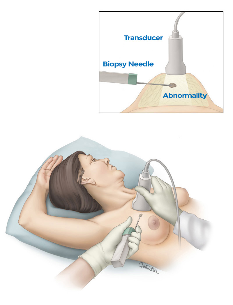
Ultrasound-Guided Breast Biopsy (UGBB) uses sound waves to help locate an abnormality and remove a sample for further examination. It is less invasive than surgical biopsy, leaves little or no scarring and does not involve exposure to ionizing radiation.
Lumps or abnormalities in the breast are often detected by physical examination, mammography, or other imaging studies. However, it is not always possible to tell from these imaging tests whether a growth is benign or cancerous. UGBB is performed to remove a small sample of cells from a suspicious area in the breast for further analysis under a microscope. Ultrasound imaging is used to help guide the radiologist’s instruments to the site of the abnormality. If the results of the analysis require that the abnormality be removed, the surgeon can use the information obtained in the biopsy to plan a course of treatment.
An ultrasound-guided breast biopsy can be performed when a breast ultrasound shows an abnormality, which could include:
- A suspicious solid mass
- A distortion in the structure of the breast tissue
- An area of abnormal tissue change
There are times when your doctor may decide that ultrasound guidance for biopsy
is appropriate even for a mass that can be felt.
How Should I Prepare for My Ultrasound-Guided Breast Biopsy?
Prior to your biopsy, you should report to your doctor any recent illnesses, allergies (especially to anesthesia) or other medical conditions, and all medications that you are currently taking, including herbal supplements. Your physician may advise you to stop taking aspirin, blood thinners, or certain herbal supplements for three to five days before your procedure.
You will be asked not to take aspirin or blood thinners for five days prior to the exam.
You should wear comfortable, loose-fitting clothing for your ultrasound exam. You will need to remove all clothing and jewelry in the area to be examined.
You may want to have a relative or friend accompany you and drive you home afterward.
What Will I Experience During the Exam?
Ultrasound scanners consist of a console containing digital electronics, a video screen,and
a transducer (a hand-held device, attached to the scanner by a cord, which sends and receives sound waves.)
When you receive the local anesthetic to numb the skin, you will feel a pin prick from the needle followed by a mild stinging sensation from the local anesthetic. The area will become numb within a few seconds. You will likely feel some pressure when the biopsy needle is inserted and during tissue sampling, which is normal.
A small amount of gel is applied to the skin to allow the sound waves to travel with minimal interference. The transducer is pressed against the skin, and sound waves are instantly measured and displayed on the monitor.
Using the images to visualize the location of the abnormality, the radiologist is able to view the biopsy needle as it advances to the location of the lesion and removes tissue samples in real-time.
You must remain very still while the imaging and biopsy are being performed. As tissue samples are taken, you may hear clicks or buzzing sounds from the sampling instrument.
During your biopsy, you will be awake and should experience little discomfort. Certain patients, including those with dense breast tissue, or abnormalities near the chest wall or behind the nipple may be more sensitive during the procedure. Most women report no scarring on the breast.
What Will I Experience After the Exam?
You may be instructed to take an over-the-counter pain reliever and to use a cold pack to minimize possible swelling or bruising. Temporary bruising is normal. You should contact your physician if you experience excessive swelling, bleeding, drainage, redness or heat in the breast.
If a marker is left inside the breast to mark the location of the biopsied lesion, it will cause no pain, disfigurement or harm. Biopsy markers are MRI compatible and will not activate metal detectors.
You should avoid strenuous activity for at least 24 hours after the biopsy. More detailed post-procedure care instructions designed specifically for you will be provided by your Radiologist.
What Are the Risks and Benefits of Ultrasound-Guided Breast Biopsy?
Ultrasound-Guided Breast Biopsy reliably provides tissue samples that can show whether a breast lump is benign or malignant. However, there is a small chance that this procedure will not provide the final answer to explain the imaging abnormality.
With UGBB it is possible to follow the motion of the biopsy needle as it moves through the breast tissue. UGBB is also able to evaluate lumps in locations which are hard to reach with other biopsy methods.
The procedure is less invasive than surgical biopsy, leaves little or no scarring and uses no ionizing radiation. Recovery time is brief and patients can soon resume their usual activities. The risk of hematoma or infection is extremely slight. Any post-procedure discomfort can be readily controlled by non-prescription pain medication.
Ultrasound-guided biopsy is less expensive than other biopsy methods.
To download this information in PDF format, click the image below…
Ultrasound-Guided Breast Biopsy is available at the following Advanced Radiology locations:
Stamford – 1259 East Main Street
Trumbull – Advanced Women’s Imaging Center – 15 Corporate Drive
Wilton – 60 Danbury Road

