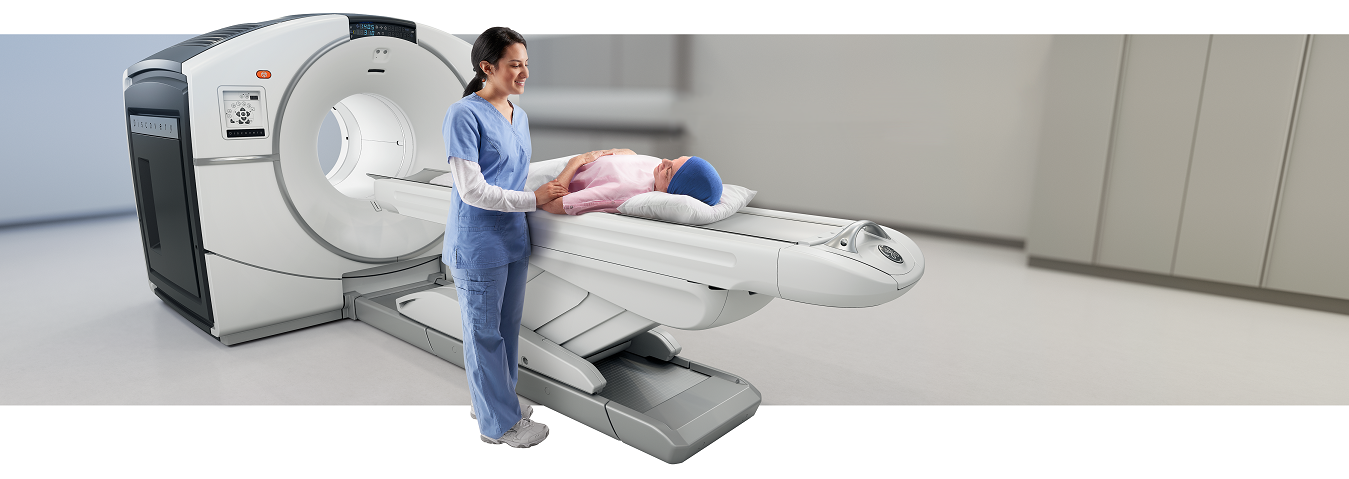
PET/CT combines Positron Emission Tomography (PET) with Computed Tomography (CT) to provide images of tissue and organ function located with precise anatomical positioning. Unlike other imaging techniques, PET/CT focus on depicting physiologic processes within the body, such as metabolism, instead of only showing anatomy.
About this ExamPreparing for this ExamRequest an AppointmentAdvanced Radiology offers Connecticut’s only permanent PET/CT scanner
at an outpatient facility
Positron Emission Tomography (PET)
Positron Emission Tomography (PET) uses small amounts of radioactive materials called radiotracers to help evaluate your organ and tissue functions, such as blood flow, sugar (glucose) metabolism, and certain cell receptors. The exam is used to diagnose a variety of diseases, including many types of cancers, heart disease, and neurological disorders such as Alzheimer’s disease.
Nuclear medicine scans, such as PET, are more sensitive than other techniques for a variety of indications, and the information gained from PET exams is often unobtainable by other imaging techniques. The PET exam results can also provide important staging and prognostic information and aid in therapy planning,
Computed Tomography (CT)
Computed Tomography (CT) imaging uses dedicated x-ray equipment to produce multiple cross-sectional images of the inside of the body.
The Benefits of Combined PET/CT Imaging
This advanced nuclear imaging technique, available at Advanced Radiology, combines Positron Emission Tomography (PET) and Computed Tomography (CT) into one machine. For the exam, detectors around the body are used to measure the amount of radiation absorption, rates of metabolism, or levels of various other chemical activity. These combined views allow the information from two different exams to be correlated and interpreted on a single image that pinpoints the anatomic location of abnormal metabolic activity within the body.
PET/CT is Commonly Used to:
- Detect cancer or determine whether a cancer has spread in the body
- Assess the effectiveness of a treatment plan, such as cancer therapy
- Determine if cancer has returned after treatment
- Evaluate blood flow to the heart muscle
- Assess the effects of a heart attack (myocardial infarction) on areas of the heart
- Identify areas of the heart muscle that would benefit from a procedure such as angioplasty or coronary artery bypass surgery (in combination with a myocardial perfusion scan)
- Evaluate brain abnormalities, such as tumors, memory disorders, seizures and other central nervous system disorders
- Map normal human brain and heart function

The combination of PET and CT scans provides information from two different exams on a single image that pinpoints the anatomic location of abnormal metabolic activity within the body.
Preparing for a PET/CT Scan
Because the radioactive tracer is effective for only a short period of time, it is important for each patient to arrive 15 minutes before their scheduled appointment and to receive the radioactive material at its scheduled time. Thus, late arrival for an appointment may require rescheduling the procedure for another day. Please note that a person who is very obese may not fit into the opening of a conventional PET/CT unit.
You will receive specific instructions based on the type of PET/CT scan you are undergoing. Diabetic patients will also receive special instructions on preparing for this exam. Test results of diabetic patients or patients who have eaten within a few hours prior to the exam can be adversely affected because of altered blood sugar or blood insulin levels.
Typically, you will be asked not to eat anything for several hours before a whole body PET/CT scan. Eating may alter the distribution of the PET tracer in your body and can lead to a suboptimal scan. This could require the scan be repeated on another day. You should not drink any liquids containing sugars or calories for several hours before the scan. Instead, you are encouraged to drink only water.
You should inform your physician and the technologist performing your exam of any medications you are taking, including vitamins and herbal supplements. You should also inform them of any allergies, especially to contrast materials, iodine, or seafood, as well as about any recent illnesses or other medical conditions.
Women should always inform their physician or technologist if there is any possibility that they are pregnant, or if they are breastfeeding. If you are breastfeeding at the time of the exam, you should ask your radiologist or referring physician how to proceed. It may help to pump breast milk ahead of time and keep it on hand for use until the radioactive tracer and contrast material are no longer in your body.
Metal objects including jewelry, eyeglasses, dentures and hair accessories should be left at home or removed prior to your exam. You may also be asked to remove hearing aids and removable dental work.
In some instances, you may be asked to change into a gown for the exam.
What Will I Experience During the Exam?
When the radiotracer is given intravenously, you will feel a slight pin prick when the needle is inserted into your vein for the intravenous line. When the radioactive material is injected into your arm, you may feel a cold sensation moving up your arm, but there are generally no other side effects. With some procedures, a catheter may be placed into your bladder, which may cause temporary discomfort.
Except for intravenous injections, most PET/CT procedures are painless and are rarely associated with significant discomfort or side effects.
Typically, it takes about one hour for the radiotracer to travel through your body and to be absorbed by the organ or tissue being studied. You will be asked to rest quietly, avoiding movement and talking. You may be asked to drink contrast material that will localize in the intestines and help the radiologist interpreting the study. You will then be moved into the PET/CT scanner. You will need to remain still during imaging. The CT scan will be done first, followed by the PET scan. On occasion, a second CT scan with intravenous contrast will follow the PET scan. The actual CT scanning takes less than two minutes. The PET scan typically takes 20-30 minutes. Total scanning time for the combined exam is approximately 30 minutes.
Total scanning time for the combined exam is approximately 30 minutes.
It is important that you remain still while the images are being recorded. Though the exam itself causes no pain, there may be some discomfort from having to remain still or to stay in one particular position during imaging. If you are claustrophobic, you may experience some anxiety while you are being scanned.
Depending on the organ or tissue is being examined, additional tests involving other tracers or drugs may be needed, which could lengthen the procedure time to as much as three hours. For example, if you are being examined for heart disease, you may undergo a PET scan both before and after exercising or before and after receiving intravenous medication that increases blood flow to the heart.
When the examination is completed, you may be asked to wait until the technologist checks the images, in case additional images are needed. Occasionally, additional images are obtained for clarification or better visualization of certain areas or structures. The need for additional images does not necessarily mean there was a problem with the exam or that something abnormal was found, and should not be a cause of concern for you.
If you had an intravenous line inserted before the procedure, it will usually be removed unless you are scheduled for an additional procedure that same day that requires an intravenous line.
What Will I Experience After the Exam?
Unless your physician tells you otherwise, you may resume your normal activities after your PET/CT scan. If any special instructions are necessary, you will be informed by a technologist, or physician before you leave the imaging facility.
Through the natural process of radioactive decay, the small amount of radiotracer in your body will lose its radioactivity during the first few hours or days following the test. You should also drink plenty of water to help flush the radioactive material out of your body as instructed by the imaging personnel.
What are the Risks and Benefits of a PET/CT Scan?
Because the dose of radiotracer administered is small, diagnostic PET/CT procedures result in relatively low radiation exposure, well within the acceptable range for diagnostic exams. Thus, the radiation risk is very low when compared to the potential benefits. There are no known long-term adverse effects from such low-dose exposure.
PET/CT examinations provide unique information, including details on both function and anatomic structure of the body that is often unattainable using other imaging procedures. For many diseases, PET/CT scans yield the most useful information needed to make a diagnosis or determine appropriate treatment.
PET/CT imaging is less expensive and may yield more precise information than exploratory surgery. By identifying changes in the body at the cellular level, imaging may detect the early onset of disease before it is evident on other imaging tests.
If you have any additional questions regarding your exam,
please call 203.337.XRAY (9729).
If you would like to schedule an appointment, click here.
PET/CT scans are available at the following Advanced Radiology locations:
Trumbull – 15 Corporate Drive

